Application of Extreme Conditions
New classes of materials have been synthesized under high pressure and temperature, exhibiting unusual physical and chemical properties, such as enhanced hardness or high-temperature superconductivity. A quite different regime of extreme conditions can be induced in solids by their interaction with heavy ions travelling at relativistic energies (GeV). These projectiles are created by high-energy particle accelerators, nuclear fission, and cosmic events and deposit exceptional amounts of kinetic energy within an exceedingly short interaction time (less than fs) into nm-sized sample volumes. Extremely high energy densities (up to tens of eV/atom) are induced, which drive the local atomic structure far from equilibrium. This results in rapid phase transition pathways through equilibrium and non-equilibrium states that trigger complex structural modifications within a highly localized nanoscale damage zone. The key strategy of our research approach is to investigate the response of materials to the coupled extreme conditions of ion irradiation (10 eV/atom), pressure (1 Mbar), and temperature (5000 K). The coupling of the extreme energy depositions of swift heavy ions with high pressure and high temperature leads to exciting applications at the interfaces of materials research, nuclear science, and geoscience. This experimental approach can be used to study the behavior of materials under extreme conditions, to form and stabilize novel phases, to manipulate the physical and chemical properties of solids at the nanoscale, and to investigate the effects of radioactive decay events in compressed and heated minerals of Earth’s interior.
 We routinely use diamond-anvil cells (DACs) to apply pressure onto samples. Pressure is created by squeezing two opposing diamond anvils against the sample chamber with an aperture (diameter 0.05 – 0.1 mm) drilled in a steel or rhenium gasket and filled with a pressure medium (e.g., methanol/ethanol). Depending on the culet face of the diamond anvils, pressures of up to a few Mbar can be reached. Resistive and laser heating can be used to simultaneously heat samples at high pressure to a few hundred to a few thousand kelvin. External heating is applied by heating wires wrapped around each diamond. In order to generate more extreme temperatures of a few thousand kelvin, DACs are coupled with intense infrared laser beams. Well-focused YAG or CO2 lasers of 50 – 100 watts of power heat the sample through one or both diamond windows. The incandescent light from the laser-heated sample is analyzed with a spectrometer through the diamond anvil, and Planck fits to the emission spectra yield the sample temperature.
 We perform irradiation experiments at large accelerator facilities using a wide range of ion species and energies. Most frequently we conduct ion-beam exposures at the GSI Helmholtz Centre for Heavy Ion Research in Darmstadt, Germany. GSI, as one of the world’s largest ion-beam facilities, is equipped with two major accelerator components: the Universal Linear Accelerator (UNILAC) and the swift-heavy ion synchrotron (SIS). Ambient-pressure irradiations are conducted at the UNILAC using typically energetic heavy ions (e.g., 2.2-GeV Au ions). High-pressure techniques have been successfully combined with the use of ion beams by injecting relativistic heavy ions (e.g., 50-GeV Au ions) from the SIS through the mm-thick diamond anvil of a high-pressure cell (M. Lang et al., Nature Materials 2009). We continuously improve the experimental protocols and are now able to simultaneously expose materials to controlled extremes of pressure (~1 Mbar) and irradiation (~1×10¹³ ions/cm2).
Materials Characterization
An important aspect of our research is the application of advanced analytical techniques, including methods that are only recently possible due to improved capabilities at large user facilities. During dedicated beamtimes, we use high-energy X-rays at synchrotron sources (e.g., Advanced Photon Source at Argonne National Laboratory) to characterize samples by means of X-ray diffraction and X-ray absorption spectroscopy (M. Lang et al., Journal of Materials Research 2015). X-ray diffraction is essential for obtaining valuable information on the nanoscale structural response of materials to irradiation or high pressure. It can be used to obtain unit-cell parameters of defective materials, establish phase fractions from irradiation- induced crystalline-to-crystalline phase transformations, investigate the amorphization process, or measure other modifications, such as changes in grain size or heterogeneous microstrain. X-ray absorption spectroscopy yields information about the electronic structure and local bonding environment of materials. This shows potential redox changes in materials after exposure to pressure or irradiation. The transparency of diamond anvils allows for in situ characterization of materials that are under pressure (and temperature).
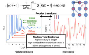
 More recently, we have included neutron total scattering experiments with pair distribution function (PDF) analysis as a new approach for the characterization of radiation effects in materials. Key to this approach is the use of GeV ions with a very high penetration depth to produce sufficiently large irradiated sample mass of about 150 mg (J. Shamblin et al., Nature Materials 2016). The Nanoscale-Ordered Materials Diffractometer (NOMAD) at the Spallation Neutron Source (Oak Ridge National Laboratory) is one of the very few neutron beamlines in the world dedicated to producing high-quality neutron total scattering data. The most important aspect of the NOMAD beamline, for our purposes, is its large range of accessible momentum transfer (Q), such that it is capable of producing PDFs with very high resolution. A PDF is obtained directly through the Fourier transform of the total scattering structure function, S(Q) – 1, from reciprocal space to real space. By including the diffuse scattering contributions, which are omitted in traditional diffraction experiments, a unique structural insight is obtained on aperiodic defect configurations including disordered and amorphous domains. Neutrons scatter strongly from low-Z elements, permitting a detailed analysis of both cation and anion defect behavior, which is particularly useful for oxide materials. PDF analysis elucidates the local defect structure, including changes in site occupation, coordination, and bond distance. It is also critical for the study of short-range structure in non-crystalline materials, as the average aperiodic atomic arrangement of glassy materials precludes the application of many other characterization techniques.
In order to explore the effect of structural modifications induced by extreme conditions on physical properties, we investigate the electric and ionic conductivity of irradiated materials. High-temperature broadband dielectric spectroscopy (also known as impedance spectroscopy) is used as an innovative method for the characterization of defect behavior and disorder in complex oxides. While many spectroscopic techniques focus on the interaction of a sample with electromagnetic waves of frequency >1012 Hz (e.g., IR- and Raman spectroscopy), broadband dielectric spectroscopy covers a wide range from 10-6 – 1012 Hz and gleans information regarding electric dipole relaxations and the translational motion of charged particles.
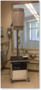
 A broadband dielectric spectrometer applies an alternating electric field to a sample and measures the impedance and current through the sample, in and out of phase, as a function of frequency. Our state-of-the-art broadband dielectric spectrometer with an integrated furnace allows us to conduct in situ measurements up to 1400 °C. An advanced gas-mixing system enables adjustment of the sample environment by mixing of three input gases (adjustable to the ppm level). Each side of the sample can be supplied with an independent gas stream, which is particularly important in separating electric and ionic components of conductivity. While broadband dielectric spectroscopy has been applied to many complex oxides during or after temperature exposure, measurements on ion-irradiated materials are not available. Similar to the proposed neutron scattering experiments, energetic ions will play a key role in extending this technique to the study of ion irradiated samples.